Lumbar Cage Introduction, Sizes, Uses and Advantages.

Lumbar Cage

Lumbar Cage Introduction

When it comes to treating lumbar spinal problems and ensuring effective fusion, the Lumbar cage has shown to be an invaluable instrument.

A Lumbar cage is a medical device used in spinal fusion surgery to treat disorders affecting the lumbar spine. It is a device that is placed into the disc space between two neighbouring vertebrae from the back.

It is intended to restore disc height, provide stability, and encourage vertebral fusion.

Lumbar Cage Sizes

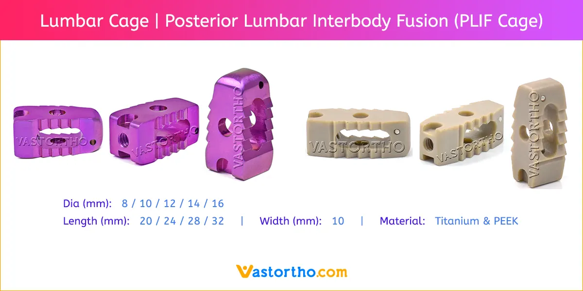
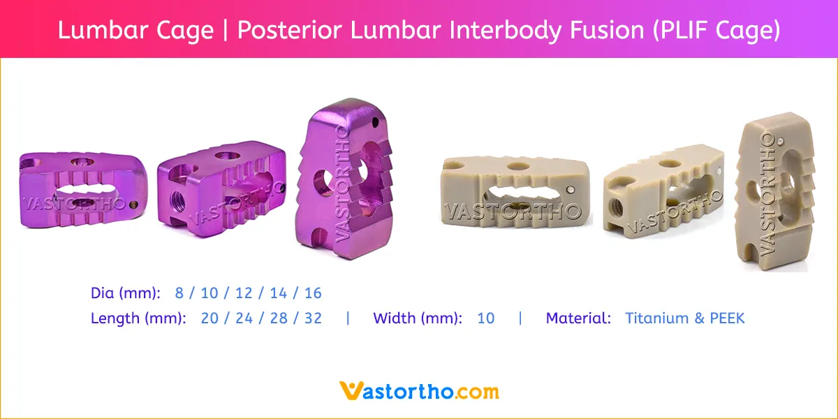
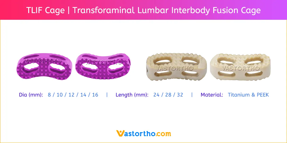
The Lumbar cage is a small, hollow device made of various materials such as titanium or polyetheretherketone (PEEK). Lumbar cages come in a variety of shapes and sizes to meet the demands of the patient.

Titanium Mesh Cage are available in various diameters and lengths.
Diameters: 8mm, 10mm, 12mm, 14mm and 16mm.
Lengths: 20mm, 24mm, 28mm and 32mm
Width: 10mm

Lumbar Cage Uses

During a PLIF procedure, the surgeon gains access to the spine by making a back incision. To reveal the spinal canal and nerve roots, the lamina (the bony arch of the vertebra) is partially removed. After that, the injured intervertebral disc is removed, making room for the Lumbar cage.

The Lumbar cage is placed in the open disc space. It can have a variety of structures, but it is often a hollow or porous device with a central channel. The cage functions as a spacer, preserving correct disc height and restoring vertebral alignment. It also serves as a platform for the placement of bone transplant material.

Lumbar Cage acts as a spacer, maintaining the disc height and restoring alignment between the vertebrae. It provides a stable environment for bone graft material to be placed, promoting fusion between the adjacent vertebrae. The fusion process involves the bone graft material growing and fusing with the adjacent vertebral bone, creating a solid, stable fusion mass.

Bone graft material can be taken from the patient’s own body (autograft) or received from a donor (allograft). The bone graft is wrapped around the Lumbar cage and encourages the formation of new bone. This causes fusion of the vertebrae over time, resulting in a solid mass that stabilises the spine.

Lumbar Cage Advantage

There are various advantages to using a Lumbar cage in spinal fusion surgery. It aids in the restoration of disc height, relieves nerve pressure, stabilises the spine, and promotes vertebral fusion. The type of Lumbar cage utilised will depend on the surgeon’s preference, the patient’s condition, and other considerations.

Stabilization: The Lumbar cage helps restore and maintain good spinal alignment by filling the intervertebral space, providing stability and minimising pain caused by aberrant motion.

Load Distribution: The cage facilitates in the distribution of load and stress over the fused segment, reducing undue strain on surrounding structures.

Increased Fusion Rates: When compared to alternative fusion procedures, the use of a Lumbar cage has been associated with better fusion rates, enhancing the likelihood of successful fusion and long-term stability.

The PLIF and TLIF techniques

Background: Posterior fusion procedures (posterior lumbar interbody fusion, PLIF; transforaminal lumbar interbody fusion, TLIF) are long-established surgical techniques for lumbar interbody fusion. They differ from anterior lumbar interbody fusion (ALIF) and extreme lateral interbody fusion (XLIF) procedures by approach and associated complications.

Objectives: The posterior fusion procedures PLIF and TLIF are presented and compared with other fusion methods, including advantages and disadvantages. Furthermore, the surgical technique and their complications are described. Based on the current literature, it is discussed which surgical techniques can be used in various cases.

Results: PLIF and TLIF procedures reduced back and leg pain, restored the sagittal profile of the lumbar spine, and achieved good fusion rates and long-term stability. Advantages of the TLIF procedure include shorter operative times, less blood loss, less intraoperative risk of injury to neural structures, and shorter convalescence. Compared with the interposition of a cage in the ALIF technique, a further step with the risk of vascular injury is eliminated.

Conclusions: The PLIF and TLIF procedures are almost equivalent posterior fusion procedures with high fusion rates, good long-term clinical outcomes, and low risk of complications. The TLIF procedure is slightly advantageous: lower nerve irritation rates, shorter operative times, and less extensive operation. Thus, the TLIF procedure is available for cases with single-sided pathologies and the PLIF procedure is available for bilateral compressions.

Do you have any questions for Lumbar Cage? Please fill this form we will get in touch with you shortly.

Name(Required)
This field is for validation purposes and should be left unchanged.

More Products from Spine Products

Lumbar Cage Contraindications

Contraindications may be qualified or total, and need to be taken into consideration when evaluating the prognosis in each case. The physician’s education, training and professional judgement must be relied upon to choose the most appropriate device and treatment. Conditions presenting an increased risk of failure include:

  • Any active or suspected latent infection or marked local inflammation in or about the affected area.
  • Compromised vascularity that would inhibit adequate blood supply to the fracture or the operative site.
  • Bone stock compromised by disease, infection or prior implantation that can not provide adequate support and/or fixation of the devices.
  • Material sensitivity, documented or suspected.
  • Obesity. An overweight or obese patient can produce loads on the implant that can lead to failure of the fixation of the device or to
    failure of the device itself.
  • Patients having inadequate tissue coverage over the operative site.
  • Implant utilization that would interfere with anatomical structures or physiological performance.
  • Any mental or neuromuscular disorder which would create an unacceptable risk of fixation failure or complications in postoperative care.
  • Other medical or surgical conditions which would preclude the potential benefit of surgery.
  • All associated diseases which could endanger the function and success of the Lumbar Cage .

Warnings and Precautionary for Lumbar Cage

Before using Lumbar Cage, the surgeon and ancillary staff should study the safety information in these instructions, as well as any product-specific information in the product description, surgical procedures and/or brochures.

Cage is made from medical grade materials and are designed, constructed and produced with utmost care. These quality Cage assure best working results provided they are used in the proper manner. Therefore, the following instructions for use and safety recommendations must be observed.

Improper use of  Cage can lead to damage to the tissue, premature wear, destruction of the instruments and injury to the operator, patients or other persons.

It is vital for the operating surgeon to take an active role in the medical management of their patients. The surgeon should thoroughly understand all aspects of the surgical procedure and instruments including their limitations. Care in appropriate selection and proper use of surgical instruments is the responsibility of the surgeon and the surgical team. Adequate surgical training should be completed before use of implants.

Factors which could impair the success of the operation:

  • Allergies to implanted materials.
  • Localized bone tumours.
  • Osteoporosis or osteomalacia.
  • System disease and metabolic disturbances.
  • Alcohol and drug abuse.
  • Physical activities involving excessive shocks, whereby the implant is exposed to blows and/or excessive loading.
  • Patients who are mentally unable to understand and comply with the doctor’s instructions.
  • Poor general health.

Possible Adverse Effects

The following adverse effects are the most common resulting from implantation:

  • Loosening of the Cage, which may result from cyclic loading of the fixation site and/or tissue reaction of the implant.
  • Early and late infection.
  • Further bone fracture resulting from unusual stress or weakened bone substance.
  • Temporary or chronic neural damage resulting from pressure or hematomata.
  • Wound hematomas and delayed wound healing.
  • Vascular disease including venal thrombosis, pulmonary embolism and cardiac arrest.
  • Heterotopic ossification.
  • Pain and discomfort due to presence of the Lumbar Cage.
  • Mechanical failure of the implant, including bending, loosening or breakage.
  • Migration of implant resulting in injury.

Preoperative Planning for Lumbar Cage

The operating planning is carried out following a thorough clinical evaluation of the patient, Also, x-rays must be taken to allow a clear indication of the bony anatomy and associated deformities. At the time of the operation, the corresponding implantation instruments in addition to a complete size of Lumbar Cage must be available.

The clinician should discuss with the patient the possible risks and complications associated with the use of implants. It is important to determine pre-operatively whether the patient is allergic to any of the implant materials. Also, the patient needs to be informed that the performance of the device cannot be guaranteed as complications can affect the life expectancy of the device.

Lumbar Cage Precautions

  • Confirm functionality of instruments and check for wear during reprocessing. Replace worn or damaged instruments prior to use.
  • It is recommended to use the instruments identified for this Cage.
  • Handle devices with care and dispose worn bone cutting instruments in a sharps container.
  • Always irrigate and apply suction for removal of debris potentially generated during implantation or removal.

Lumbar Cage Warnings

  • Lumbar Cage can break during use (when subjected to excessive forces). While the surgeon must make the final decision on removal of the broken part based on associated risk in doing so, we recommend that whenever possible and practical for the individual patient, the broken part should be removed. Be aware that implants are not as strong as native bone. Implants subjected to substantial loads may fail.
  • Instruments, screws and cut plates may have sharp edges or moving joints that may pinch or tear user’s glove or skin.
  • Take care to remove all fragments that are not fixated during the surgery.
  • While the surgeon must make the final decision on implant removal, we recommend that whenever possible and practical for the individual patient, fixation devices should be removed once their service as an aid to healing is accomplished. Cage removal should be followed by adequate post-operative management to avoid refracture.

Lumbar Cage General Adverse Events

As with all major surgical procedures, risks, side effects and adverse events can occur. While many possible reactions may occur, some of the most common include: Problems resulting from anesthesia and patient positioning (e.g. nausea, vomiting, dental injuries, neurological impairments, etc.), thrombosis, embolism, infection, nerve and/or tooth root damage or injury of other critical structures including blood vessels, excessive bleeding, damage to soft tissues incl. swelling, abnormal scar formation, functional impairment of the musculoskeletal system, pain, discomfort or abnormal sensation due to the presence of the device, allergy or hypersensitivity reactions, side effects associated with hardware prominence, loosening, bending, or breakage of the device, mal-union, non-union or delayed union which may lead to breakage of the Lumbar Cage, reoperation.

PLIF with cages and local bone graft in the treatment of spinal stenosis

Posterior lumbar interbody fusion (PLIF) implants are increasingly being used for 360 degrees fusion after decompression of lumbar spinal stenosis combined with degenerative instability. Both titanium and PEEK (PolyEtherEtherKetone) implants are commonly used. Assessing the clinical and radiological results as well as typical complications, such as migration of the cages, is important. In addition, questions such as which radiological parameters can be used to assess successful fusion, and whether the exclusive use of local bone graft is sufficient, are frequently debated. We prospectively evaluated 30 patients after PLIF instrumentation for degenerative lumbar spinal canal stenosis, over a course of 42 months. In all cases, titanium cages and local bone graft were used for spondylodesis. The follow-up protocol of these 30 cases included standardised clinical and radiological evaluation at 3, 6, 12 and 42 months after surgery. Overall satisfactory results were achieved. With one exception, a stable result was achieved with restoration of the intervertebral space in the anterior column. After 42 months of follow-up in most cases, a radiologically visible loss of disc space height can be demonstrated. Clinically relevant migration of the cage in the dorsal direction was detected in one case. Based on our experience, posterior lumbar interbody fusion (PLIF) can be recommended for the treatment of monosegmental and bisegmental spinal stenosis, with or without segmental instability. Postoperative evaluation is mainly based on clinical parameters since the titanium implant affects the diagnostic value of imaging studies and is responsible for artefacts. The results observed in our group of patients suggest that local autologous bone graft procured from the posterior elements after decompression is an adequate material for bone grafting in this procedure.

PLIF using local facet joint autograft and pedicle screw fixation

Study design: This is a retrospective study of 42 patients having lumbar degenerative disease or spondylolytic spondylolisthesis treated by posterior lumbar interbody fusion (PLIF) using local autogenous facet joint graft and pedicle screw fixation with an average follow-up time of 8.5 years.

Objectives: To evaluate the radiographic and clinical results of patients treated with PLIF using adjacent facet joint autograft and pedicle screw internal fixation.

Summary of background data: Some goals of spinal surgery have been achieved by interbody arthrodesis using a posterior approach popularized by Cloward. However, significant problems including bone graft collapse, resorption, nonunion, persistent neurologic compression, and iliac crest donor complication using the classic PLIF remain. There are few reports describing the results of a PLIF by total facet joint excision.

Methods: Forty-two patients (average, 53.2 years) treated at our institution with PLIF by total facetectomy were followed for an average period of 8.5 years. The changes in the Japanese Orthopedic Association score, the recovery rate, complications, and radiographic findings were evaluated.

Results: Good radiographic fusion (92.9%) and clinical results (postoperative recovery rate of 76% in the Japanese Orthopedic Association score) were achieved by PLIF using local facet joint autograft and pedicle screw fixation in treating patients with debilitating lumbar degenerative disease. The complications related to the operative procedure occurred in three patients of delayed union.

Conclusions: For lumbar degenerative diseases with osteophytic changes of facet joints, PLIF using pedicle screw fixation and local autogenous bones obtained from facet excision may be justified as a treatment opinion. The procedure as described offers advantages for spinal surgery when PLIF is warranted.