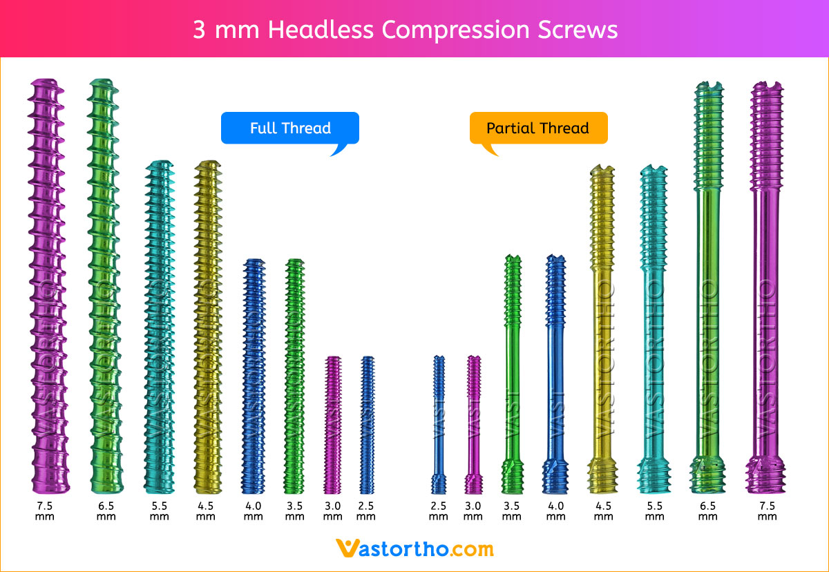
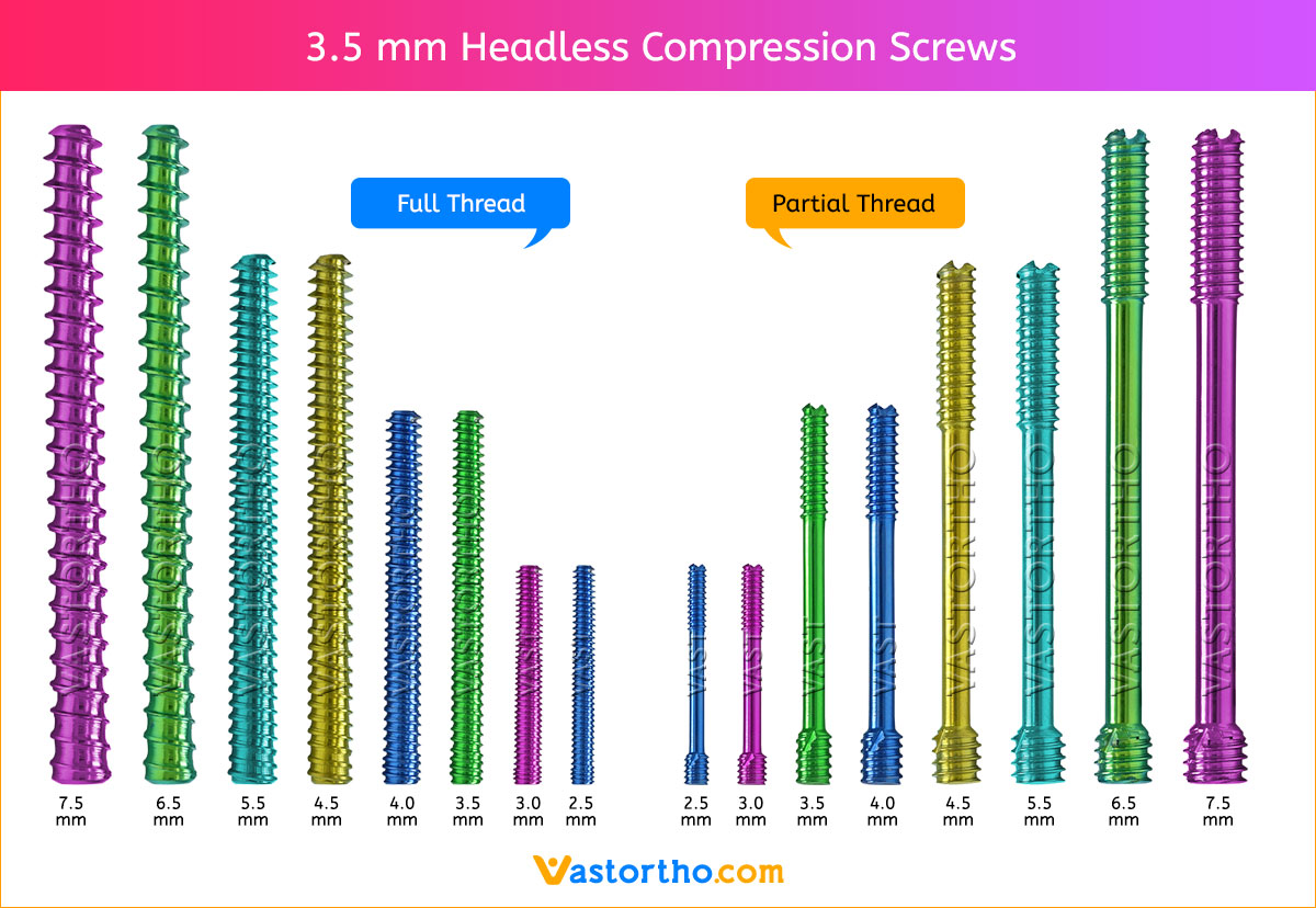
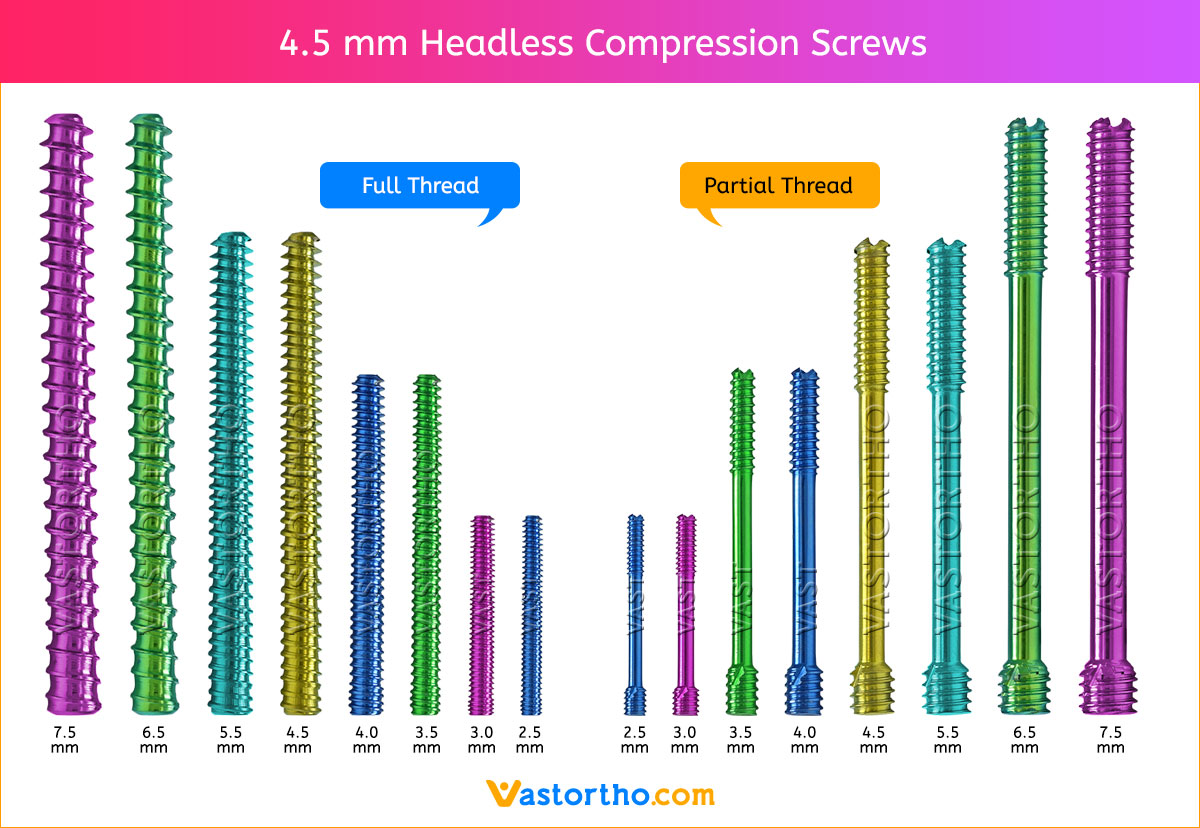
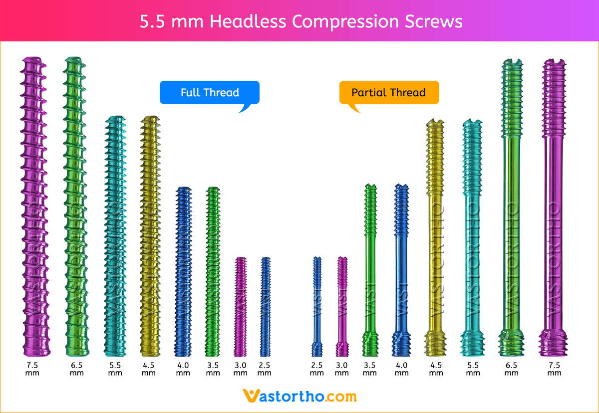
Headless Compression Screws

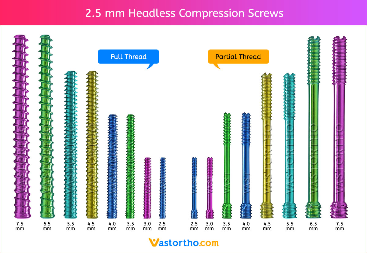
2.5 mm Headless Compression Screws
3 mm Headless Compression Screws
3.5 mm Headless Compression Screws
4 mm Headless Compression Screws
4.5 mm Headless Compression Screws
5.5 mm Headless Compression Screws
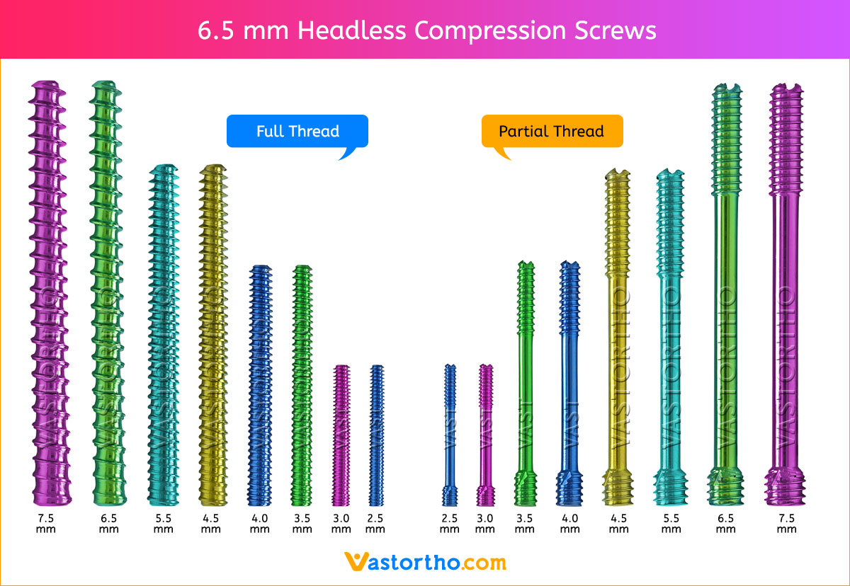
6.5 mm Headless Compression Screws
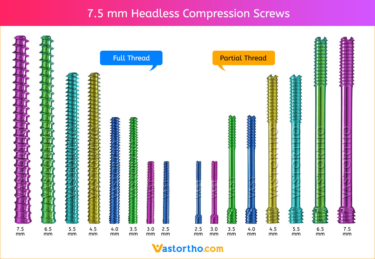
7.5 mm Headless Compression Screws

Headless Compression Screws Specification

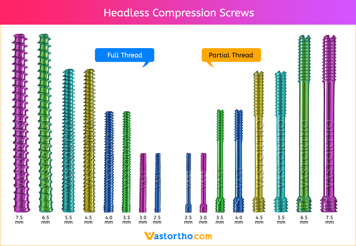
  • Headless design: Headless design allows the screw implantation in and around articular regions with minimal soft tissue irritation or risk of interference.
  • Cannulated, for accurate insertion: Cannulated design facilitates accurate percutaneous insertion with minimal soft tissue damage.
  • Gradual compression through variable thread pitch: The wider thread pitch at the tip of the screw penetrates the bone faster than the finer trailing threads, compressing the two fragments gradually as the screw is advanced.
  • Two types of screws, Fully threaded, Partially threaded.
  • Fully threaded: Fully threaded screw provides a higher holding force resulting in increasing stability.
  • Sharp cutting flutes: Sharp cutting flutes in the screw tip facilitate screw insertion.
  • Headless Compression Screws available in both Titanium and Stainless Steel.
  • Sterile and non-sterile packaging options available for Headless Compression Screws.

Headless Compression Screws

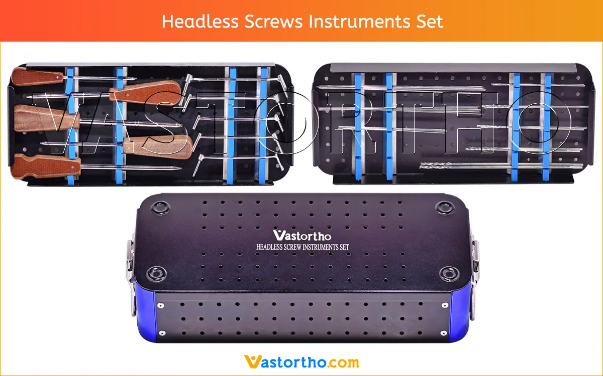
Headless Compression Screws Instruments Set

Headless Screws Instruments Set Image

A complete instruments set are available for Headless Compression Screws. Instruments can be modified according to the customer’s requirement with minimum quantity required. All these instruments can be used several times.

We are keeping wide range of  instruments items in this set to ensures that Doctors get almost all required items during Surgery. Below is list of items of this set.

  • Cannulated Screw Driver, 1.5 mm Tip – 1 Qty
  • Cannulated Screw Driver, 2.0 mm Tip – 1 Qty
  • Cannulated Screw Driver, 2.5 mm Tip – 1 Qty
  • Cannulated Screw Driver, 3.0 mm Tip – 1 Qty
  • Cannulated Screw Driver, 3.5 mm Tip – 1 Qty
  • Cannulated Drill Bit, 2 mm – 2 Qty
  • Cannulated Drill Bit, 2.5 mm – 2 Qty
  • Cannulated Drill Bit, 2.7 mm – 2 Qty
  • Cannulated Drill Bit, 3.2 mm – 2 Qty
  • Cannulated Drill Bit, 4 mm – 2 Qty
  • Double Drill Guide, 2 x 0.8 mm – 1 Qty
  • Double Drill Guide, 2.5 x 1 mm – 1 Qty
  • Double Drill Guide, 2.7 x 1.2 mm – 1 Qty
  • Double Drill Guide, 3.2 x 1.1 mm – 1 Qty
  • Double Drill Guide, 4 x 1.8 mm – 1 Qty
  • Guide Wire, 0.8 mm – 10 Qty
  • Guide Wire, 1.2 mm – 10 Qty
  • Guide Wire, 1.8 mm – 5 Qty
  • Graphics Aluminum Box with Silicone Fittings – 1 Qty

Headless Compression Screws Uses

Headless Compression Screws Uses

Headless Compression Screws Uses 2

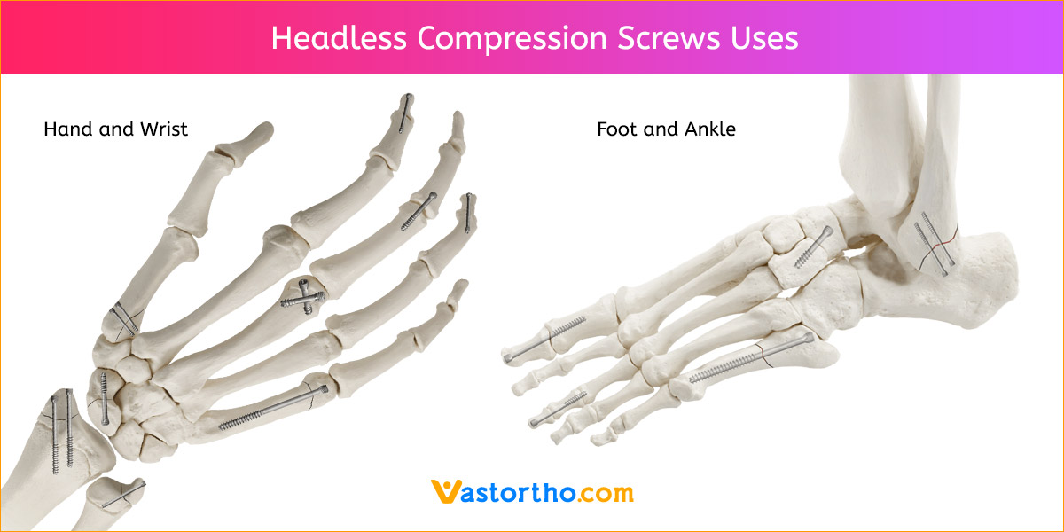
Headless Compression Screws are designed to be used in bone reconstruction, osteotomy, arthrodesis, joint fusion, fracture repair and fracture fixation of bones appropriate for the size of the device.

Headless Compression Screws are intended for bone fixation of the hand and foot flowing trauma or osteotomy. Self ­Tapping and reverse cutting flutes on both ends of the screw aid with insertion and removal. Tapered profile gaining compression and maximizing pull-out strength along its entire length.

Headless Compression Screws 2.5 mm, 3 mm, 3.5 mm, 4 mm, 4.5 mm, and 5.5 mm are intended for use as fixation devices for small bones, bone fragments, and osteotomies. They are not intended for interference or soft tissue fixation.

Headless Compression Screws 6.5 mm and 7.5 may be used for fusions, fractures, or osteotomies of the clavicle, humerus, radius, ulna, ilium, femur, patella, fibula, tibia, talus, malleolus, and calcaneus.

Headless Compression Screws Sizes

2.5 mm Screws Length

8mm, 10mm, 12mm, 14mm, 16mm, 18mm, 20mm, 22mm, 24mm, 26mm, 28mm and 30mm.

3 mm Screws Length

8mm, 10mm, 12mm, 14mm, 16mm, 18mm, 20mm, 22mm, 24mm, 26mm, 28mm and 30mm.

3.5 mm Screws Length

8mm, 10mm, 12mm, 14mm, 16mm, 18mm, 20mm, 22mm, 24mm, 26mm, 28mm, 30mm, 32mm, 34mm, 36mm, 38mm, 40mm, 42mm, 44mm, 46mm, 48mm, 50mm, 55mm, 60mm, 65mm, 70mm, 75mm and 80mm.

4 mm Screws Length

8mm, 10mm, 12mm, 14mm, 16mm, 18mm, 20mm, 22mm, 24mm, 26mm, 28mm, 30mm, 32mm, 34mm, 36mm, 38mm, 40mm, 42mm, 44mm, 46mm, 48mm, 50mm, 55mm, 60mm, 65mm, 70mm, 75mm and 80mm.

4.5 mm Screws Length

8mm, 10mm, 12mm, 14mm, 16mm, 18mm, 20mm, 22mm, 24mm, 26mm, 28mm, 30mm, 32mm, 34mm, 36mm, 38mm, 40mm, 42mm, 44mm, 46mm, 48mm, 50mm, 55mm, 60mm, 65mm, 70mm, 75mm and 80mm.

5.5 mm Screws Length

12mm, 14mm, 16mm, 18mm, 20mm, 22mm, 24mm, 26mm, 28mm, 30mm, 32mm, 34mm, 36mm, 38mm, 40mm, 42mm, 44mm, 46mm, 48mm, 50mm, 55mm, 60mm, 65mm, 70mm, 75mm, 80mm, 85mm, 90mm, 95mm, 100mm, 105mm, 110mm, 115mm and 120mm.
6.5 mm Screws Length

12mm, 14mm, 16mm, 18mm, 20mm, 22mm, 24mm, 26mm, 28mm, 30mm, 32mm, 34mm, 36mm, 38mm, 40mm, 42mm, 44mm, 46mm, 48mm, 50mm, 55mm, 60mm, 65mm, 70mm, 75mm, 80mm, 85mm, 90mm, 95mm, 100mm, 105mm, 110mm, 115mm and 120mm.

7.5 mm Screws Length

12mm, 14mm, 16mm, 18mm, 20mm, 22mm, 24mm, 26mm, 28mm, 30mm, 32mm, 34mm, 36mm, 38mm, 40mm, 42mm, 44mm, 46mm, 48mm, 50mm, 55mm, 60mm, 65mm, 70mm, 75mm, 80mm, 85mm, 90mm, 95mm, 100mm, 105mm, 110mm, 115mm and 120mm.

Headless Compression Screws Surgical Techniques

Volar Scaphoid Technique for 2.5 mm, 3 mm, 3.5 mm and 4 mm Headless Compression screws.

1. Approach and Needle Insertion

The procedure can be carried out using the volar traction approach or using a conventional volar type approach with the arm supine on a hand table. The volar traction approach facilitates reduction of a displaced fracture and permits arthroscopy to ensure accuracy of the reduction.
Fluoroscopy is used throughout.

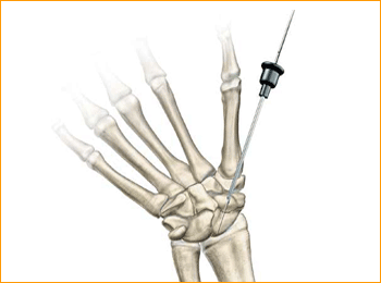
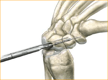
The entry point is then located using a 12 or 14 gauge IV needle introduced on the antero-radial aspect of the wrist just radial to and distal to the scaphoid tuberosity. This serves as a trochar for the guide wire and is a directional aid to establish a central path along the scaphoid. The needle is then insinuated into the scaphotrapezial joint, tilted into a more vertical position and the position is checked on the under image
intensifier. By gently levering on the trapezium this maneuver brings the distal pole of the scaphoid more radial and thus ultimately facilitates screw insertion. The entry point should be approximately 1/3 the way across the scaphoid from the tuberosity in the A/P plane and central in the lateral plane.

2. Guide Wire Insertion

Pass the guide wire through the needle and drill it across the fracture, continually checking the direction on the image intensifier and correcting as necessary, aiming for the radial aspect of the proximal pole. It is extremely important not to bend the guide wire and any adjustments in direction should be made using the needle as a guide rather than attempting to alter the line of the guide wire alone.

3. Determine Screw Length

Advance the guide wire to stop just short of the articular surface as the wire should not breach it at this stage. The position, alignment and length are checked once more. Make a simple stab incision at the entry point of the wire, and deepen this down to the distal pole of the
scaphoid using a small hemostat and blunt dissection.

Determine the length of the screw either with the appropriate depth gauge or by advancing a second guide wire of the same length up the distal cortex of the scaphoid and subtracting the difference between the two. When using the volar approach, the correct screw size is 2–4 mm shorter than the measured length so as to ensure that the proximal tip of the screw is fully buried below the cartilage and the cortical surface.

4. Advance Guide Wire

Advance the guide wire through the proximal pole of the scaphoid so as to exit on the dorsal aspect of the wrist. This is a precautionary measure to minimize the risk of inadvertent withdrawal of the wire during the reaming process and screw insertion and to facilitate removal of the
proximal portion if the wire were to break. A second de-rotation wire can then be inserted in those cases where it is felt that there is a possibility of rotational instability of the fracture.

5. Drill

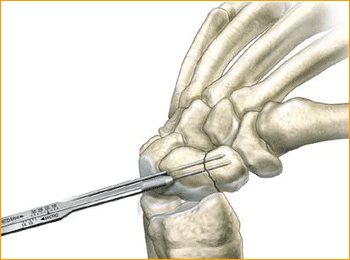
Remove the 12 gauge needle and pass the Cannulated Profile Drill over the wire using either a power drill or by hand stopping 1–2 mm short of the articular surface. The long drill is recommended to mitigate the effects of varying bone density and distraction upon screw insertion.

6. Advance Self-Tapping Screw

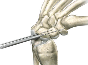
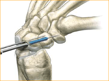
The self-tapping screw is then advanced over the guide wire and the wire removed. Compression can then be confirmed radiographically on the image intensifier.
1. Approach and Needle Insertion

Headless Compression Screws Surgical Techniques 1

2. Guide Wire Insertion

Headless Compression Screws Surgical Techniques 2

3. Determine Screw Length

Headless Compression Screws Surgical Techniques 3

4. Advance Guide Wire

Headless Compression Screws Surgical Techniques 4

5. Drill

Headless Compression Screws Surgical Techniques 5

6. Advance Self-Tapping Screw

Headless Compression Screws Surgical Techniques 6

Dorsal Scaphoid Technique for 2.5 mm, 3 mm, 3.5 mm and 4 mm Headless Compression screws.

1.   APPROACH AND NEEDLE INSERTION
The entry point in the proximal pole is at the tip of the scaphoid immediately adjacent to the scapholunate ligament. This can be located either using an arthroscopy or mini open dorsal approach between the third and fourth extensor compartments. Whichever approach is employed, it is essential to ensure that the guide wire does not transfix an extensor tendon.

Having established the entry point, introduce the appropriate guide wire aiming for the base of the thumb and check the position on the fluoroscope. Aim to place the leading edge of the guide wire in the subchondral surface of the distal pole of the scaphoid. Confirm the wire placement and depth under imaging.

2.  FRACTURE STABILIZATION
If the fracture is unstable it may be helpful to place a second parallel guide wire using the parallel wire guides.

3.  DETERMINE SCREW LENGTH
Measure guide wire length using either the percutaneous screw sizer, or by placing a second wire at the entry point and subtracting the difference. The screw sizer cannot be used with the arthroscopic technique due to the limited access. Subtract 4 mm from the measured length to ensure that both ends of the screw are buried within the bone.

4.  ADVANCE GUIDE WIRE
Advance the guide wire through the far cortex so that it lies in the subcutaneous tissues. This minimizes the risk of accidental withdrawal of the guide wire while drilling and facilitates wire removal if it should break.
Tip: For most adult males the screw should not be longer than 26 mm, and in females 22 mm.

5.  DRILL NEAR CORTEX
Open the near cortex with the appropriate profile drill.

6.  DRILL FAR FRAGMENT
Next, drill into the far fragment with the long drill. To be effective the drill only has to advance 4–5 mm past the fracture site.
Tip: The long drill is recommended to mitigate the effects of varying bone density and distraction upon screw insertion.

7.  SCREW INSERTION
Insert the correctly sized screw with the appropriate hex driver. If resistance is met upon insertion or if distraction occurs, stop, remove the screw, redrill with the long drill, and re-insert the screw. Confirm placement and length of the screw on imaging, ensuring that both leading and trailing edges of the screw are beneath the articular surfaces. Finally remove the guide wires.

1. Approach and Needle Insertion

Headless Compression Screws Surgical Techniques 7

2. Fracture Stabilization

Headless Compression Screws Surgical Techniques 8

3. Determine Screw Length

Headless Compression Screws Surgical Techniques 9

4. Advance Guide Wire

Headless Compression Screws Surgical Techniques 10

5. Drill Near Cortex

Headless Compression Screws Surgical Techniques 11

6. Drill Far Fragment

Headless Compression Screws Surgical Techniques 12

7. Screw Insertion

Headless Compression Screws Surgical Techniques 13

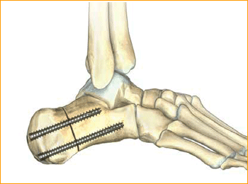
Jones Fracture Technique for 4.5 mm and 5.5 mm Headless Compression screws.

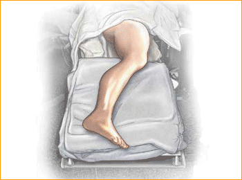
1.  PATIENT POSITIONING
Position the patient in a semi-lateral position utilizing a bean bag body positioner. The patient should be moved to the distal end of the bed and the operative leg draped free as the side up. Exertion of the operative limb should be checked prior to prep and drape to confirm that
the operative limb can be positioned on the mini c-arm during surgery.

2.  INDICATION AREA OUTLINE
The base of the fifth metatarsal is outlined, including the insertions of the peroneus brevis and tertius tendons.

3.  APPROACH AND EXPOSURE
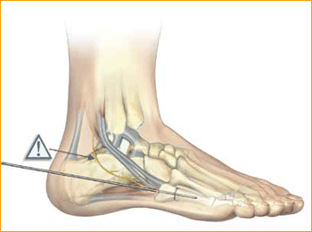
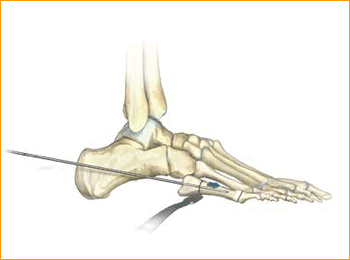
The guide wire, .062″, for the 4.7 Screw can be positioned at the base of the fifth metatarsal under fluoroscopic guidance. A small incision is made at the base of the fifth metatarsal at the intersection of the peroneus brevis and tertius tendons. Care is made to identify and protect the sural nerve branches which run over the peroneal tendons. If necessary, fibers of the lateral aponeurosis and peroneus brevis tendon are separated and retracted away from the styloid process of the base of the fifth metatarsal. A mini Hohman Retractor is placed on the plantar
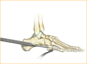
aspect of the base of the fifth metatarsal. The surgeon’s fingers can be used to reduce the fifth metatarsal fracture by placing them in between the fourth and fifth metatarsals. This closes down the fifth metatarsal fracture site during guide wire, drill, and screw placement. A guide wire is drilled from the base of the fifth metatarsal into the central portion of the metatarsal shaft. It is maintained within the intramedullary canal in order to avoid distal penetration. Confirm placement with fluoroscopy.

4.  MEASURE DEPTH
Depth is measured from the exposed portion of the guide wire with the cannulated depth gauge.

5.  ADVANCE GUIDE WIRE
After selecting the size, advance the guide wire approximately 5 mm to maintain distal pin fixation before drilling.
Caution: Make sure not to compromise distal joint surfaces when advancing the guide wire.

6.  SOFT TISSUE GUIDE PLACEMENT
Place the soft tissue guide (the guide should be used throughout) over the guide wire and open the near cortex using the appropriate cannulated profile drill.

7.  DRILL
Leaving the soft tissue guide in place, drill into the far fragment with the appropriate cannulated, long drill. Reference the markings on the drill to confirm desired depth.
Tip: The long drill is recommended to mitigate the effects of varying bone density and distraction upon screw insertion.

8.  FRACTURE COMPRESSION
In order to account for countersinking and fracture compression, a screw that measures 5 mm shorter than the measured total depth is inserted over the guide wire while protecting the soft tissues with a soft tissue guide.

9.  SCREW INSERTION
The screw is placed while under fluoroscopic guidance in order to avoid cortical penetration. Postoperative protocol: The patient is placed into a soft dressing, supported by a fiberglass splint. Patients can be made non-weight-bearing for a period of 2–6 weeks postoperatively depending upon Torg type of fracture, bone quality, and underlying morbidities.

1. Patient Positioning

Headless Compression Screws Surgical Techniques 14

2. Indication Area Outline

Headless Compression Screws Surgical Techniques 15

3. Approach and Exposure

Headless Compression Screws Surgical Techniques 16

4. Measure Depth

Headless Compression Screws Surgical Techniques 17

5. Advance Guide Wire

Headless Compression Screws Surgical Techniques 18

6. Soft Tissue Guide Placement

Headless Compression Screws Surgical Techniques 19

7. Drill

Headless Compression Screws Surgical Techniques 20

8. Fracture Compression

Headless Compression Screws Surgical Techniques 21

9. Screw Insertion

Headless Compression Screws Surgical Techniques 22

Calcaneal Osteotomy Technique for 6.5 mm Headless Compression screws.

1.   PATIENT POSITIONING
Position the patient at the end of the bed, semi-lateral. Check that the leg can be placed easily onto the mini c-arm prior to preparation of the operative limb.

2.  APPROACH AND EXPOSURE
An incision is made posterior to the peroneal tendons, perpendicular to the body of the calcaneus. Cephalad and caudal mini Hohman Retractors are
placed to protect the neurovascular structures and plantar fascia. Care is made to preserve the peroneal tendons and the sural nerve.

3.  CREATE OSTEOTOMY
An oscillating saw is used to make the osteotomy cut perpendicular to the body of the calcaneus. The saw is not used to complete the cut through the medial cortex. This is completed with an osteotome in order to avoid damaging medial neurovascular structures.

4.  GUIDE WIRE PLACEMENT
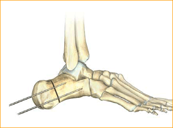
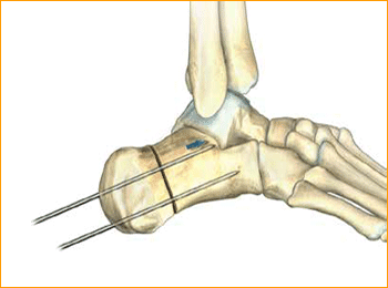
The body of the calcaneus is displaced medially or laterally and held in place with two guide pins. The distal portion of the pins are placed at the volar aspect of the angle of Gissane in order to capture solid bone distally and assist with compression of the osteotomy by the screws.
Confirm guide pin placement under fluoroscopy.
Tip: The soft tissue protector and arthroscopic probe can be used to assist in guide wire placement.

5.  MEASURE DEPTH
Select a screw the same size as measured. However, to account for countersinking and compression it is common to select a screw one size shorter
than the measured depth. After selecting the size, advance the guide wire approximately 5 mm to maintain distal pin fixation before drilling.
Caution: Make sure not to compromise joint surfaces when advancing the guide wire.

6.  SELECT SCREW SIZE
Place the soft tissue guide (the guide should be used throughout) over the guide wire and open the near cortex using the appropriate cannulated profile drill.

7.  SOFT TISSUE GUIDE
Place the soft tissue guide over the guide wire and open the near cortex using the appropriate cannulated profile drill.
Tip: Drills should be advanced slowly with continuous irrigation to minimize the potential of heat build-up. Clean drill periodically during each procedure to optimize performance.

8.  DRILL
Leaving the soft tissue guide in place, drill into the far fragment with the appropriate cannulated, long profile drill. Reference the markings on the drill to confirm desired depth.
Tip: The long drill is recommended to mitigate the effects of varying bone density and distraction upon screw insertion.

9.  SCREW INSERTION
Cannulated  Screws are placed. Placement is confirmed by lateral and axial fluoroscopy views in the operating room.
Caution: The marking on the driver shows when the screw is approximately flush with the end of the soft tissue protector; assure that the soft tissue protector is touching bone to accurately determine screw depth. Verify final screw position with fluoroscopy.

10.  ADDITIONAL SCREW PLACEMENT
Repeat steps 5–9 for each additional screw placement. The operative limb is placed into a bulky compression dressing. A splint is also placed. Patients are made non-weight-bearing in a cast, boot, or splint for 6 weeks after surgery.

1. Patient Positioning

Headless Compression Screws Surgical Techniques 23

2. Approach and Exposure

Headless Compression Screws Surgical Techniques 24

3. Create Osteotomy

Headless Compression Screws Surgical Techniques 25

4. Guide Wire Placement

Headless Compression Screws Surgical Techniques 26

5. Measure Depth

Headless Compression Screws Surgical Techniques 27

6. Select Screw Size

Headless Compression Screws Surgical Techniques 28

7. Soft Tissue Guide

Headless Compression Screws Surgical Techniques 29

8. Drill

Headless Compression Screws Surgical Techniques 30

9. Screw Insertion

Headless Compression Screws Surgical Techniques 31

10. Additional Screw Placement

Headless Compression Screws Surgical Techniques 32

Headless Compression Screws Contraindications

Contraindications may be qualified or total, and need to be taken into consideration when evaluating the prognosis in each case. The physician’s education, training and professional judgement must be relied upon to choose the most appropriate device and treatment. Conditions presenting an increased risk of failure include:

  • Any active or suspected latent infection or marked local inflammation in or about the affected area.
  • Compromised vascularity that would inhibit adequate blood supply to the fracture or the operative site.
  • Bone stock compromised by disease, infection or prior implantation that can not provide adequate support and/or fixation of the devices.
  • Material sensitivity, documented or suspected.
  • Obesity. An overweight or obese patient can produce loads on the implant that can lead to failure of the fixation of the device or to
    failure of the device itself.
  • Patients having inadequate tissue coverage over the operative site.
  • Implant utilization that would interfere with anatomical structures or physiological performance.
  • Any mental or neuromuscular disorder which would create an unacceptable risk of fixation failure or complications in postoperative care.
  • Other medical or surgical conditions which would preclude the potential benefit of surgery.
  • All associated diseases which could endanger the function and success of the Headless Compression Screws.

Warnings and Precautionary for Headless Compression Screws

Before using Headless Compression Screws, the surgeon and ancillary staff should study the safety information in these instructions, as well as any product-specific information in the product description, surgical procedures and/or brochures.

Headless Compression Screws is made from medical grade materials and are designed, constructed and produced with utmost care. These quality Screws assure best working results provided they are used in the proper manner. Therefore, the following instructions for use and safety recommendations must be observed.

Improper use of  Screws can lead to damage to the tissue, premature wear, destruction of the instruments and injury to the operator, patients or other persons.

It is vital for the operating surgeon to take an active role in the medical management of their patients. The surgeon should thoroughly understand all aspects of the surgical procedure and instruments including their limitations. Care in appropriate selection and proper use of surgical instruments is the responsibility of the surgeon and the surgical team. Adequate surgical training should be completed before use of implants.

Factors which could impair the success of the operation:

  • Allergies to implanted materials.
  • Localized bone tumours.
  • Osteoporosis or osteomalacia.
  • System disease and metabolic disturbances.
  • Alcohol and drug abuse.
  • Physical activities involving excessive shocks, whereby the implant is exposed to blows and/or excessive loading.
  • Patients who are mentally unable to understand and comply with the doctor’s instructions.
  • Poor general health.

Possible Adverse Effects

The following adverse effects are the most common resulting from implantation:

  • Loosening of the Screws, which may result from cyclic loading of the fixation site and/or tissue reaction of the implant.
  • Early and late infection.
  • Further bone fracture resulting from unusual stress or weakened bone substance.
  • Temporary or chronic neural damage resulting from pressure or hematomata.
  • Wound hematomas and delayed wound healing.
  • Vascular disease including venal thrombosis, pulmonary embolism and cardiac arrest.
  • Heterotopic ossification.
  • Pain and discomfort due to presence of the Screws.
  • Mechanical failure of the implant, including bending, loosening or breakage.
  • Migration of implant resulting in injury.

Preoperative Planning for Headless Compression Screws

The operating planning is carried out following a thorough clinical evaluation of the patient, Also, x-rays must be taken to allow a clear indication of the bony anatomy and associated deformities. At the time of the operation, the corresponding implantation instruments in addition to a complete size of Headless Compression Screws must be available.

The clinician should discuss with the patient the possible risks and complications associated with the use of implants. It is important to determine pre-operatively whether the patient is allergic to any of the implant materials. Also, the patient needs to be informed that the performance of the device cannot be guaranteed as complications can affect the life expectancy of the device.

Headless Compression Screws Precautions

  • Confirm functionality of instruments and check for wear during reprocessing. Replace worn or damaged instruments prior to use.
  • It is recommended to use the instruments identified for this Headless Compression Screws.
  • Handle devices with care and dispose worn bone cutting instruments in a sharps container.
  • Always irrigate and apply suction for removal of debris potentially generated during implantation or removal.

Headless Compression Screws Warnings

  • Headless Compression Screws can break during use (when subjected to excessive forces). While the surgeon must make the final decision on removal of the broken part based on associated risk in doing so, we recommend that whenever possible and practical for the individual patient, the broken part should be removed. Be aware that implants are not as strong as native bone. Implants subjected to substantial loads may fail.
  • Instruments, screws and cut plates may have sharp edges or moving joints that may pinch or tear user’s glove or skin.
  • Take care to remove all fragments that are not fixated during the surgery.
  • While the surgeon must make the final decision on implant removal, we recommend that whenever possible and practical for the individual patient, fixation devices should be removed once their service as an aid to healing is accomplished. Headless Screws removal should be followed by adequate post-operative management to avoid refracture.

Headless Compression Screws General Adverse Events

As with all major surgical procedures, risks, side effects and adverse events can occur. While many possible reactions may occur, some of the most common include: Problems resulting from anesthesia and patient positioning (e.g. nausea, vomiting, dental injuries, neurological impairments, etc.), thrombosis, embolism, infection, nerve and/or tooth root damage or injury of other critical structures including blood vessels, excessive bleeding, damage to soft tissues incl. swelling, abnormal scar formation, functional impairment of the musculoskeletal system, pain, discomfort or abnormal sensation due to the presence of the device, allergy or hypersensitivity reactions, side effects associated with hardware prominence, loosening, bending, or breakage of the device, mal-union, non-union or delayed union which may lead to breakage of the Headless Compression Screws, reoperation.

Bone screws are the most commonly used orthopedic implants. There are many different types and sizes of screws for different types of bones. Most bone screws are made out of stainless steel or titanium alloys. The outer diameter, root diameter, and thread pitch and angle are important in determining screw mechanics.

In orthopedics, screws are typically described by their outer diameter, for example, a “5.5 mm Headless Compression Screws” has an outside diameter of 5.5 mm. The pitch of a screw is the linear distance travelled by a screw for one full turn of the screw. The screw advances by a distance equal to the distance between the threads with each full turn. Cortical screws have a lower pitch and therefore more number of threads. Cancellous bone screws have a greater depth of the screw to increase the surface area and therefore improve the purchase, as the bone is weaker.

Screws function by converting the tightening torque into internal tension in the screw and elastic reactions in the surrounding bone. This creates compression between the fracture fragments that the screw is holding together. 2mm Cortical Screw is typically inserted into holes drilled equal to the root diameter and are either self-tapping or are inserted tapped (threaded) holes. The torque to insert cortical bone screws can be high, so the screws must be properly inserted into the correct size drilled hole and designed to withstand insertion torque levels expected in cortical bone. Cancellous bone screws have large, deep threads that grip the spongy bone well. Because of the relatively low strength of the cancellous bone, failure of the screw itself during insertion is rare, but pull out can be an issue.